The usage of proteins is almost inevitable in most biochemical experiments. The ironic thing is, even if several billion or trillion proteins are present right in front of us, we never really get to see their true form due to their microscopic sizes. Thus, I enrolled in a class named structural biology, which I learned four programs: PyMOL, Swiss PDB Viewer, MolMol, and Chimera, for visualizing proteins, their physical properties, and several interaction mechanisms. This helped me understand important structural properties about the protein I had been studying.
Here I demonstrate some simulation methods implemented on the protein: signal transducer and activator of transcription 3 (STAT3). Three PDB files are used in this project: 3cwg, 1bg1, and 1bf5.
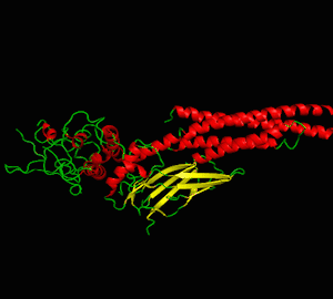
Analysis 1: Protein visualization using cartoon (top-left), dots (top-right), sticks (bottom-left) and spheres (bottom-right). Secondary structures such as the alpha helix and beta sheet are colored differently (PDB ID: 3cwg) (PyMOL).


Analysis 2: The volume of the protein (PDB ID: 1bg1) is calculated as 71.613nm3, and its surface area is calculated as 263.23nm2. The structure is transformed into a spherical molecular representation prior to calculation (Chimera).
Analysis 3: Total width and height of the protein (PDB ID: 3cwg) (Swiss PDB Viewer).

Analysis 4: Morphing between two different PDB files of the same protein (PDB ID: 3cwg and 1bg1) (Chimera). The blue structure is 3cwg, and the gray-white structure is 1bg1 in the lower figure.


Analysis 5: Electric charge on alpha helix (PDB ID: 3cwg) (PyMol).

Analysis 6: Mutation of Proline to Histidine at residue 255 (PDB ID: 3cwg) (Swiss PDB Viewer).


Analysis 7: Twisting of the ϕ and ψ angle (respectively left and right figure) at residue 255 (Proline) (PDB ID: 3cwg) (Swiss PDB Viewer).

ϕ angle 
ψ angle
Analysis 8: Ramachandran plots of the same protein with two different PDB files (PDB ID: 3cwg (left figure) and 1bg1 (right figure)) (MolMol).

3cwg 
1bg1
Analysis 9: Coulomb force on protein surface. The surface is colored from red (-10kcal/mol×e) to blue (10kcal/mol×e) gradient in order to indicate differences in Coulombic forces (PDB ID: 3cwg) (Chimera).
Analysis 10: The hydrogen bond between the two SH2 domains of the STAT3 dimer (PDB ID: 1bg1) (Chimera).

Analysis 11: Morphing between STAT3 (1bg1) and STAT1 (1bf5) (another similar protein of the STAT family) (Chimera). The blue structure is STAT1, and the white structure is STAT3 in the lower figure.

